Xenium In Situ
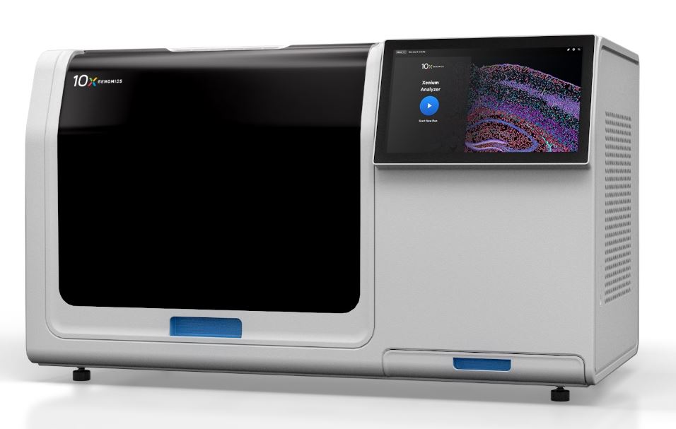
Xenium by 10x Genomics is a state-of-the-art single cell spatial imaging platform. It integrates high-resolution imaging and onboard data analysis to profile 100s-1000s of RNA targets in FFPE or FF tissue samples using either pre-designed (Human or Mouse) or customized gene expression panels. Xenium offers best-in class sensitivity, specificity and high confident transcript-to-cell assignments using multimodal cell segmentation.
You can find more information about Xenium by visiting 10x Genomics Xenium Resource Portal
Here you can find Products, Brochures & Flyers, Spatial Tech Notes, Presentations, Workflow Illustrations, Customized Panel Information and Data Showcase of Images.
How to get started with a Xenium project in the Bioimaging Center:
- To get started with a Xenium project, contact a Bioimaging staff member to schedule a project consultation
- Sample Type – Human or Mouse panels available. Contact 10x Genomics if you are interested in custom panels
- Tissue Compatibility
- Formalin-Fixed Paraffin-Embedded (FFPE) tissue (5um thickness)
- Fresh Frozen (FF) tissue (10um thickness)
- Total sample area on slides – 10.45mm x 22.45mm
- Maximum of 2 slides per Xenium run
Data Analysis:
There are several tools that can be used for data analysis. Some include Xenium Explorer, Xenium Ranger and Third-Party Tools. For Xenium Explorer, a designated computer workstation can be reserved in the Bioimaging Center for data processing.
Location: APB 141Q

Zeiss PALM MicroBeam Laser Capture Microdissection Microscope
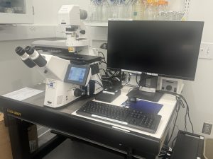
The Zeiss PALM MicroBeam Laser Capture Microdissection (LCM) Microscope is a dual use system.
- As an LCM, it uses a pulsed solid-state 355nm (UV-A) laser for isolating specific cells for downstream analysis. It can isolate DNA, RNA, proteins, chromosomes, single cells, small organisms and live cells. Samples for LCM can be viewed in either brightfield or fluorescence mode. The LCM can isolate samples on slides or in culture dishes. Supporting substrates can be either standard glass microscope slides or special membrane slides (PEN or PET slides). PET membrane slides are ideal for fluorescence work. The LCM is compatible with both cryostat-sectioned or formalin fixed and paraffin embedded tissue. The Palm Robo software is used for LCM collection.
- The system can also be utilized as an inverted microscope for both brightfield and fluorescent imaging. Advanced imaging techniques such as tile sets, Z stacks and time series are capable using the ZEN Blue software.
Cameras:
- Color: Axiocam 503 color
- Fluorescent: Axiocam 503 mono
Operating Software:
- PALM Robo 4.8
- ZEN Blue 2.6 Pro
- AxioVision SE64 4.9.1
Fluorescent Filter Sets:
- Blue/DAPI
- Green/FITC
- Red/TRITC
- Far Red/Cy5
Objectives:
- 5x Fluar/0.25
- 10x Fluar/0.5, DIC II
- 20x LD PlanNeofluar/0.4, DIC II
- 40x LD PlanNeofluar/0.6, DIC II
- 63x LD PlanNeofluar/0.75, DIC II
- 100x PlanNeofluar/1.3, Oil, DIC III
Location: APB 141Q

