Staff

Dr. Sylvain Le Marchand
Zeiss 880 Confocal Training and Appointments
Phone: (302) 831-3441
sylvain@udel.edu
Office: AB 141M

Jean Ross
Cryostat Training
Phone: (302) 831-0162
jeanross@udel.edu
Office: APB 141L

APB 141J
Deborah Powell
Cryostat Training
Phone: (302) 831-3449
dpowell@udel.edu
Office: APB 141J
Equipment
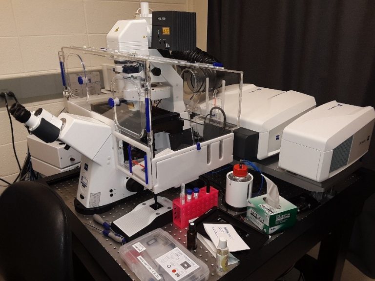
Zeiss 880 at Wolf Hall - Confocal / Spectral / Airyscan

The LSM 880 microscope is a fully automated multimodal microscope that can be used for confocal and spectral imaging. The Airyscan detector enables super-resolution and high speed imaging.
The LSM880 is fully equipped for live-cell imaging. It has an environmental enclosure (temperature, humidity, and CO2 control) and z-drift correction to maintain samples at the focal plane over time.
Applications:
- Confocal imaging.
- Spectral imaging with linear unmixing. It can be used to remove background autofluorescence or for multicolor imaging when spectral overlaps between colors cannot be separated with standard confocal microscopy.
- Super-resolution imaging. The Airyscan can provide a 2-fold increase in resolution compared to conventional confocal microscopy.
- High-speed imaging. The Airyscan detector can be used to image 4 times faster than the confocal mode (up to 20fps at 512*512). The 880 is also equipped with a piezo-electric stage for high-speed acquisition of z-stacks.
- Confocal reflection microscopy. It can be used to image collagen fibers and surface microtopography.
- FRET, FRAP, FLIP.
- Fluorescence Correlation Spectroscopy imaging. It provides quantitative data on concentrations, diffusion coefficients, molecular transport and interactions.
- Tile scanning to image a large field at high resolution.
- Live cell imaging thanks to the environmental enclosure.
Objectives: 5x/0.25, 10x/0.3, 20x/0.8, 40x/1.2 W, 40x/1.3 Oil, 63x/1.4 Oil.
Laser lines: 405nm, 458nm, 488nm, 514nm, 561nm, 594nm, 633nm.
Detectors: a 32-channel ultra-sensitive spectral GaAsP PMT (Gallium Arsenide Phosphide PhotoMultiplier Tube) detector (i.e. ChS in Zen) which is ideal for spectral imaging, two multi-alkali PMT detectors (Ch1 & Ch2), one transmitted PMT (T-PMT) for contrast imaging (Bright Field, Differential Interference Contrast, Phase), and one GaAsP Airyscan detector for super-resolution and fast imaging.
Operating software: Zen 2.3 SP1
Location: Wolf Hall #304
Contact person: Dr. Le Marchand
Leica 3050 Cryostat at Wolf Hall
The Leica CM3050 cryostat is used to cut sections of frozen samples embedded in a tissue freezing medium.
Location: Wolf Hall #267
Contact persons: Deborah Powell or Jean Ross
