Zeiss LSM880 at APBio - Confocal / Spectral / Multiphoton / Airyscan imaging

The LSM880 microscope is a fully automated multimodal microscope that can be used for confocal, spectral, and multiphoton excitation imaging. The Airyscan detector enables super-resolution and high speed imaging.
The LSM880 is fully equipped for live-cell imaging. It has an environmental enclosure (temperature, humidity, and CO2 control) and z-drift correction to maintain samples at the focal plane over time.
Applications:
- Confocal imaging.
- Spectral imaging with linear unmixing. It can be used to remove background autofluorescence or for multicolor imaging when spectral overlaps between colors cannot be separated with standard confocal microscopy.
- Multiphoton excitation microscopy. It can be used to image up to 10x deeper than confocal microscopy.
- Super-resolution imaging. The Airyscan can provide a 2-fold increase in resolution compared to conventional confocal microscopy.
- High-speed imaging. The Airyscan detector can be used to image 4 times faster than the confocal mode (up to 20fps at 512x512). The LSM880 is also equipped with a piezo-electric stage for high-speed acquisition of z-stacks.
- Confocal reflection microscopy. It can be used to image collagen fibers and surface microtopography.
- FRET, FRAP, FLIP.
- Fluorescence Correlation Spectroscopy imaging. It provides quantitative data on concentrations, diffusion coefficients, molecular transport and interactions.
- Tile scanning to image a large field at high resolution.
- Live cell imaging thanks to the environmental enclosure.
Objectives: 10x/0.3, 20x/0.8, 40x/1.2 W, 40x/1.3 Oil, 63x/1.4 Oil – more objectives are available as add-ons.
Laser lines: 405nm, 458nm, 488nm, 514nm, 561nm, 633nm, 680-1080nm.
Detectors: a 32-channel ultra-sensitive spectral GaAsP PMT (Gallium Arsenide Phosphide PhotoMultiplier Tube) detector (i.e. ChS in Zen) which is ideal for spectral imaging, two multi-alkali PMT detectors (Ch1 & Ch2), one transmitted PMT (T-PMT) for contrast imaging (Bright Field, Differential Interference Contrast, Phase), and one GaAsP Airyscan detector for super-resolution and fast imaging.
Operating software: Zen 2.3 SP1
Location: APB 141S
Contact person: Dr. Sylvain Le Marchand
Zeiss LSM880 at Wolf Hall - Confocal / Spectral / Airyscan imaging

The LSM880 microscope is a fully automated multimodal microscope that can be used for confocal and spectral imaging. The Airyscan detector enables super-resolution and high speed imaging.
The LSM880 is fully equipped for live-cell imaging. It has an environmental enclosure (temperature, humidity, and CO2 control) and z-drift correction to maintain samples at the focal plane over time.
Applications:
- Confocal imaging.
- Spectral imaging with linear unmixing. It can be used to remove background autofluorescence or for multicolor imaging when spectral overlaps between colors cannot be separated with standard confocal microscopy.
- Super-resolution imaging. The Airyscan can provide a 2-fold increase in resolution compared to conventional confocal microscopy.
- High-speed imaging. The Airyscan detector can be used to image 4 times faster than the confocal mode (up to 20fps at 512x512). The 880 is also equipped with a piezo-electric stage for high-speed acquisition of z-stacks.
- Confocal reflection microscopy. It can be used to image collagen fibers and surface microtopography.
- FRET, FRAP, FLIP.
- Fluorescence Correlation Spectroscopy imaging. It provides quantitative data on concentrations, diffusion coefficients, molecular transport and interactions.
- Tile scanning to image a large field at high resolution.
- Live cell imaging thanks to the environmental enclosure.
Objectives: 5x/0.25, 10x/0.3, 20x/0.8, 40x/1.2 W, 40x/1.3 Oil, 63x/1.4 Oil.
Laser lines: 405nm, 458nm, 488nm, 514nm, 561nm, 594nm, 633nm.
Detectors: a 32-channel ultra-sensitive spectral GaAsP PMT (Gallium Arsenide Phosphide PhotoMultiplier Tube) detector (i.e. ChS in Zen) which is ideal for spectral imaging, two multi-alkali PMT detectors (Ch1 & Ch2), one transmitted PMT (T-PMT) for contrast imaging (Bright Field, Differential Interference Contrast, Phase), and one GaAsP Airyscan detector for super-resolution and fast imaging.
Operating software: Zen 2.3 SP1
Location: Wolf Hall #304
Contact person: Dr. Sylvain Le Marchand
Zeiss LSM980 at CBBI - Confocal / Spectral / Multiphoton imaging
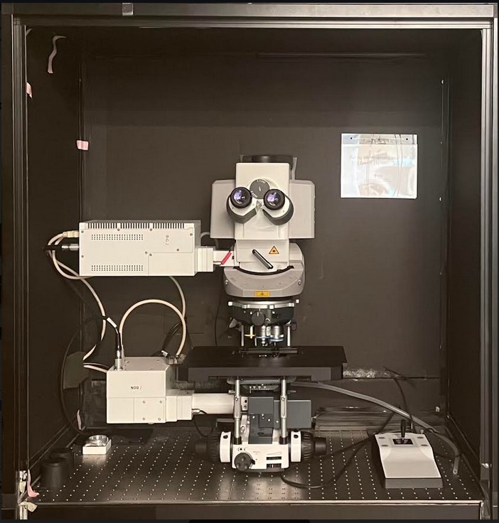
The Zeiss LSM980 is an upright confocal microscope (i.e. objectives are above the sample) that can be used for confocal, spectral, and multiphoton imaging.
Applications:
- Confocal imaging.
- Spectral imaging with linear unmixing. It can be used to remove background autofluorescence or for multicolor imaging when spectral overlaps between colors cannot be separated with standard confocal microscopy.
- Multiphoton excitation microscopy. It can be used to image up to 10x deeper than confocal microscopy. Two non-descanned PMT detectors can be used for imaging multiphoton fluorescence or second harmonic generation (SHG) in the forward and reverse directions. SHG is now widely used to image collagen in many different applications, and is becoming useful for imaging myosin and some polysaccharides.
- Confocal reflection microscopy. It can be used to image collagen fibers and surface microtopography.
FRET, FRAP, FLIP. - Fluorescence Correlation Spectroscopy imaging. It provides quantitative data on concentrations, diffusion coefficients, molecular transport and interactions.
- Tile scanning to image a large field at high resolution.
Objectives: 10x/0.3, 20x/0.75, 40x/1.2 W, 40x/1.4 Oil – more objectives are available as add-ons.
Laser lines: 405nm, 488nm, 561nm, 639nm, 690-1040nm.
Detectors: a 32-channel ultra-sensitive spectral GaAsP PMT (Gallium Arsenide Phosphide PhotoMultiplier Tube) detector (i.e. ChS in Zen) which is ideal for spectral imaging, two GaAsP PMT detectors (Ch1 & Ch2), one transmitted PMT (T-PMT) for contrast imaging (Bright Field, Oblique illumination, Differential Interference Contrast), and two non-descanned detectors (one GaAsP and one multi-alkali) for multiphoton imaging.
Operating software: Zen 3.5
Location: CBBI 244C
Contact person: Dr. Sylvain Le Marchand
Zeiss CellDiscoverer7 at APBio – Confocal / High Content Screening / Airyscan imaging

The Celldiscoverer 7 is an automated boxed microscope with a user-friendly interface that allows wide-field, confocal (LSM 900), and super-resolution imaging by means of its Airyscan2 detector. The system is an inverted microscope capable of live cell imaging experiments with a variety of incubation conditions. It has enhanced automated functionality such as Autocorr objectives, which enable the system to automatically detect a variety of sample holders and adjust for spherical aberrations. The CD7 is also equipped with an autoimmersion system that automatically applies water to its water immersion lenses and maintains the volume of immersion water to permit live cell imaging over a long duration. The system uses LED illumination, which has low phototoxicity and long-term stability.
Applications:
- Live cell imaging
- High-throughput imaging
- Tiling
- Time-lapse
- Optical sectioning for 3D imaging
- Super resolution imaging with the Airyscan 2
- FRAP and FRET
Objectives:
- PlanApochromat 5x/0.12
- PlanApochromat 20x/0.7 and 20X/0.95, Autocorr
- PlanApochromat 50x/1.2 water, Autocorr and autoimmersion
These objectives are used in combination with a 0.5X, 1x and 2x optovar lens, which permits 2.5x, 5x, 10x, 20x, 25x, 40x, 50x, 100x magnifications.
Laser Lines: 405 nm, 488 nm, 561 nm, and 640 nm
Detectors:
- For LSM900 Confocal:
- Airyscan 2 detector
- 2 PMT detectors
- For Wide-field Imaging:
- Axiocam 506 black and white camera (2752 × 2208 pixels)
Stage Inserts:
- The system accommodates most multiwell plates, 3.5 and 6 cm petri dishes, microscopy slides, and chamber slides
Operating Software: Zen Blue
Location: APB 141T
Contact Person: Dr. Chandran Sabanayagam
NIIMBL Stellaris 8 at APBio – Confocal / TauSTED / FLIM imaging
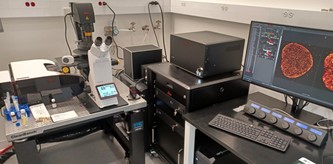
The NIIMBL Stellaris 8 tauSTED/FLIM Confocal Microscope is a fully automated multimodal microscope that can be used for confocal, fluorescence lifetime imaging microscopy (FLIM) and super-resolution (STED).
Applications:
- Confocal imaging of up to 5 colors simultaneously (far red dye with excitation and emission above 700nm can be imaged).
- FLIM can be used to separate multiple overlapping fluorophores or remove background signal. FLIM can also detect changes in the molecular environments of fluorophores and can be sensitive to multiple biomedical processes including disease progression and drug efficacy. FLIM can be used to follow fast molecular interactions via FLIM-FRET (Förster Resonance Energy Transfer) and FLIM biosensors can detect changes in metabolic state and microenvironment.
- 2D or 3D stimulated emission depletion (STED) is a super-resolution microscopy technique capable of improving spatial resolution by a factor of ~8 over standard confocal microscopy.
Objectives: 20x/0.75 multi-immersion (water, glycerol, or oil), 40x/1.1 W MotCorr, 86x/1.2W MotCorr, 93x/1.3 Glyc MotCorr, 100x/1.4 Oil – a 10x/0.4 dry is also available upon request.
MotCorr objectives can be used to compensate mismatch of refractive indexes and restore optimal imaging conditions particularly when imaging deeper into the samples.
Laser lines: 405nm, white light laser (WLL) 440-790nm, 592nm STED, 775nm STED.
The WLL is a tunable laser – one can select any laser lines between 440 and 790nm.
Detectors: HyD S1, HyD X2, HyD S3, HyD X4, HyD R5.
HyD detectors are 2 to 3 times more sensitive than regular photomultiplier detectors:
Operating software: LAS X
Location: APB 141U
Contact Person: Dr. Sylvain Le Marchand
Andor Dragonfly at APBio- Spinning Disk Confocal / TIRF / Super Resolution
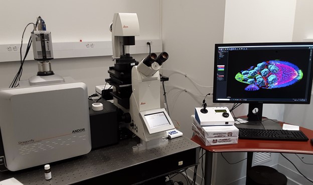
The Dragonfly microscope is a fully automated multimodal microscope that can be used for confocal, total internal reflection microscopy (TIRF) and super-resolution (localization-based techniques such as: PALM, dSTORM, PAINT, etc.).
Applications:
- Spinning disk confocal imaging. In contrast to point scanning confocals, such as the LSM880 and CD7 that serially acquire one focal point at a time to reconstitute a 2D image, the Dragonfly acquires confocal images by simultaneously acquiring thousands of spots from the focal plane. This setup enables the Dragonfly to provide several important advantages over point-scanning confocals:
- Faster speed (up to ~200 frames per second), which is ideal for fast 2D or 3D time-series or tile scanning.
- Gentler imaging – photobleaching and phototoxicity are greatly reduced, which is better for live cell imaging.
- The high sensitivity of cameras is well suited for dim specimens.
- Super-resolution imaging. The Dragonfly can perform 3D localization- based microscopy techniques (dSTORM, PAINT, etc.) that improve spatial resolution to ~30nm laterally, and ~50nm axially.
- TIRF microscopy. a technique allowing the confined excitation of fluorophores within ~100nm above the coverslip.
Objectives: 10x/0.45, 20x/0.8, 25x/0.95 W, 40x/1.1 W, 63x/1.47 Oil, 100x/1.47 Oil
Laser lines: 405nm, 445nm, 488nm, 515nm, 561nm, 638nm, 735nm.
Detectors: Two Andor Zyla 4.2 sCMOS cameras can be used together to acquire two colors simultaneously.
Operating software: Fusion
Location: APB 141R
Contact Persons: Dr. Sylvain Le Marchand (confocal), Tim Chaya (TIRF and super-resolution)
Andor Dragonfly at Wolf Hall- Spinning Disk Confocal / TIRF

The Dragonfly microscope is a fully automated microscope that can be used for confocal and total internal reflection microscopy (TIRF).
Applications:
- Spinning disk confocal imaging. In contrast to point scanning confocals, such as the LSM880 and CD7 that serially acquire one focal point at a time to reconstitute a 2D image, the Dragonfly acquires confocal images by simultaneously acquiring thousands of spots from the focal plane. This setup enables the Dragonfly to provide several important advantages over point-scanning confocals:
- Faster speed (up to ~200 frames per second), which is ideal for fast 2D or 3D time-series or tile scanning.
- Gentler imaging – photobleaching and phototoxicity are greatly reduced, which is better for live cell imaging.
- The high sensitivity of the camera is well suited for dim specimens.
- TIRF microscopy. a technique allowing the confined excitation of fluorophores within ~100nm above the coverslip.
Objectives: 1.25x/0.04, 10x/0.45, 20x/0.8, 40x/1.1 W, 63x/1.47 Oil.
Laser lines: 405nm, 488nm, 561nm, 637nm.
Detectors: Andor Sona sCMOS
Operating software: Fusion
Location: Wolf Hall #304
Contact Persons: Dr. Sylvain Le Marchand (confocal), Tim Chaya (TIRF)